hourlong learnings #4: motor unit recruitment during resistance training
A skeletal muscle is controlled through motor units. Each motor unit contains one lower motor neuron and the muscle fibers supplied by its axon. The CNS changes muscle force by recruiting more units and changing how frequently recruited units fire.
A rough model is:
$$ \text{descending + sensory input} \rightarrow \text{alpha motor-neuron spikes} \rightarrow \text{muscle-fiber action potentials} \rightarrow \text{cross-bridge force} \rightarrow \text{joint torque} $$
Recruitment, muscle force, surface EMG amplitude, and perceived effort are related mathematically, but they aren’t the same measurement.
1. what a motor unit physically contains
A motor unit contains:
alpha motor-neuron cell body in the spinal cord or brainstem
motor axon leaving through a peripheral nerve
axon branches and neuromuscular junctions
every skeletal-muscle fiber innervated by that neuron
An action potential reaching the motor terminal opens voltage-gated calcium channels. Calcium entry causes acetylcholine release. Acetylcholine binds nicotinic receptors on the motor end plate, producing an end-plate potential. If that depolarization reaches threshold, a muscle-fiber action potential travels along the sarcolemma and T-tubules. Calcium is then released from the sarcoplasmic reticulum and allows actin-myosin cross-bridge cycling.
The neural action potential is all-or-none. Motor-unit force is not. Force depends on how often the neuron fires, how many fibers it innervates, fiber cross-sectional area, muscle length, shortening velocity, calcium handling, and fatigue.
Small motor units usually innervate fewer, slow, fatigue-resistant fibers. Large units usually innervate more fibers that produce greater and faster force but fatigue sooner. Extra-ocular and hand muscles need small force increments, while large muscles like the quadriceps can use much larger units or groups of them.
2. recruitment and rate coding are separate controls
recruitment means adding another motor unit to the active pool.
Rate coding means changing the discharge frequency of a unit that is already active.
A motor-unit spike train can be written as:
$$ s_i(t) = \sum_{k=1}^{K_i} \delta(t-t_{ik}) $$
$t_{ik}$ is the time of spike $k$ from motor unit $i$. Its mean discharge rate over a window $\Delta t$ is:
$$ f_i = \frac{K_i}{\Delta t} \quad \mathrm{Hz} $$
If $h_i(t)$ is the twitch-force response to one spike, total muscle force can be approximated by:
$$ F(t) = \sum_{i=1}^{N} r_i(t) \left[s_i(t)*h_i(t)\right] $$
$r_i(t)$ is 1 when unit $i$ is recruited and 0 when it is not. $*$ is convolution. Closely spaced spikes summate because the next twitch begins before the prior twitch has relaxed. At sufficiently high firing rates, separate twitches approach fused tetanic force.
The following diagram is useful because it separates neural excitation, muscle activation, contraction dynamics, and measured force. EMG sits near the excitation side of this chain; a force plate or dynamometer sits at the mechanical end.

3. why smaller units are usually recruited first
During a gradual voluntary contraction, motor units are generally recruited from low threshold to high threshold. This is Henneman’s size principle.
For motor neuron $i$, a basic membrane approximation is:
$$ \Delta V_i \approx R_{\mathrm{in},i}I_{\mathrm{syn},i} $$
$R_{\mathrm{in},i}$ is input resistance and $I_{\mathrm{syn},i}$ is net synaptic current. Recruitment occurs when:
$$ V_{\mathrm{rest},i} + R_{\mathrm{in},i}I_{\mathrm{syn},i}(t) \geq V_{\mathrm{th},i} $$
Smaller motor neurons have higher input resistance, so the same synaptic current produces a larger voltage change. They reach spike threshold before larger neurons.
| Motor-unit group | Threshold | Twitch force | Contraction speed | Fatigue resistance |
|---|---|---|---|---|
| slow | low | low | slow | high |
| fast fatigue-resistant | middle | moderate | fast | moderate-high |
| fast fatigable | high | high | fast | low |
Synaptic noise, task, contraction speed, fatigue, sensory input, pain, and neurologic disease can change thresholds. However, the general low-to-high threshold order still holds better than the gymbro claim that someone can voluntarily choose fast-twitch fibers while leaving low-threshold units off.
4. heavy loads and fatigued light loads reach high-threshold units differently
a heavy repetition requires high force immediately, so high-threshold units are recruited early. A light first repetition can be completed with a smaller active pool. During repeated contractions, active fibers produce less force and the CNS must increase drive to maintain the same external output.
Let current force capacity fall with fatigue fraction $\phi(t)$:
$$ F_{\mathrm{available}}(t) = F_0\left[1-\phi(t)\right] $$
Relative demand is:
$$ D(t) = \frac{F_{\mathrm{required}}(t)} {F_{\mathrm{available}}(t)} $$
If required force remains $300$ N while available force falls from $1{,}000$ N to $400$ N, demand rises from $0.30$ to $0.75$. The load didn’t change, but recruitment, rate coding, and perceived effort must increase overall.
Low-load work near failure can therefore recruit high-threshold units. It does not follow that a light set and heavy set create identical firing rates, fiber force, joint stress, velocity, or training adaptation. Failure is simply the point where the system can no longer satisfy the task under the required technique and ROM.
5. fatigue can be peripheral or central
Peripheral fatigue occurs at or distal to the neuromuscular junction. Relevant mechanisms include reduced membrane excitability, altered sodium-potassium gradients, metabolite accumulation, and sarcoplasmic-reticulum calcium release.
Central fatigue is a failure to maintain sufficient voluntary neural drive. Group III and IV muscle respond to mechanical and metabolic conditions and can reduce spinal motoneuron output. Pain, hyperthermia, sleep loss, illness, and some medications can also change voluntary drive.
Let $A(t)$ be muscle activation and $F_{\max}(t)$ be current force capacity:
$$ F(t) = A(t)F_{\max}(t) $$
To maintain force $F_{\mathrm{req}}$:
$$ A_{\mathrm{req}}(t) = \frac{F_{\mathrm{req}}} {F_{\max}(t)} $$
As $F_{\max}$ falls, required activation rises. Once activation and compensation are insufficient, bar velocity falls or the repetition stops.
6. compound lifts raise whole-body effort through several systems
A squat, deadlift, or row uses multiple joints, prime movers, synergists, antagonists, stabilizers, grip, trunk bracing, and ventilation. A single-joint exercise reduces the number of major contributors.
compound-lift RPE:
local muscle effort + bracing + balance + grip + ventilation + technique

single-joint RPE:
more strongly influenced by one local muscle group
A deadlift can feel like RPE 9 because of spinal loading, grip, bracing, and total motor command without proving maximal recruitment in one hamstring region. A leg extension can create high local quadriceps effort with less cardiorespiratory and stabilizing demand.
RIR is an estimate of remaining repetitions:
$$ RIR = N_{\mathrm{failure}} – N_{\mathrm{completed}} $$
Velocity loss provides another measurement:
$$ VL = 1- \frac{v_{\mathrm{last}}} {v_{\mathrm{fastest}}} $$
If the fastest repetition is $0.60$ m/s and the last is $0.36$ m/s, velocity loss is $40%$. Neither RIR nor velocity loss directly identifies the recruited units, but both are more specific than saying a set felt hard.
7. chemical compounds can change output without matching changes in RPE
Perception of effort is strongly related to a corollary discharge of central motor command. A simple approximation is:
$$ e(t) \propto \frac{u(t)}{u_{\max}} $$
$u(t)$ is current motor command and $u_{\max}$ is the command associated with maximal voluntary effort under that condition. This is not a clinical RPE formula. It states that a larger required command usually feels harder. Pain, ventilation, temperature, expectation, and afferent feedback can change pacing and the command needed to continue.
Caffeine antagonizes adenosine receptors and can alter arousal, pain, vigilance, and central drive. A 2021 resistance-training meta-analysis reported about $0.87$ additional bench-press repetitions and a $2.01$ kg improvement in maximal strength, but no reliable reduction in RPE. A 2025 meta-analysis reported standardized mean differences of $0.42$ for mean bar velocity and $0.21$ for mean power.
The forest plot below shows the caffeine effect on mean resistance-exercise velocity. The pooled effect favors caffeine, but heterogeneity is high, so we can’t be absolutely conclusive. The medical point is that increased output does not require an equal reduction in perceived effort.

Creatine affects a different step through the creatine-kinase reaction:
$$ PCr+ADP+H^+ \rightleftharpoons Cr+ATP $$
Increasing the phosphocreatine pool supports rapid ATP resynthesis during repeated high-intensity work. It doesn’t selectively turn on high-threshold motor units. Beta-alanine increases muscle carnosine, while sodium bicarbonate increases extracellular buffering. These compounds are more relevant to repeated glycolytic work than to recruitment during one maximal repetition.
Analgesics and stronger stimulants can reduce pain or raise willingness to continue without reducing tissue stress. Using pain reduction as proof of safe loading is fundamentally weak because pain, effort, tissue force, blood pressure, and temperature can change in their own ways too.
8. disease changes recruitment in different ways
Motor-unit testing is clinically useful because weakness can originate at several levels:
upper motor neuron: cortex or corticospinal tract
lower motor neuron: anterior horn cell, root, plexus, or peripheral motor axon
neuromuscular junction: transmission from axon terminal to muscle
muscle fiber: membrane, contractile, metabolic, or inflammatory disease
In lower motor-neuron loss, surviving axons can sprout collateral branches and re-innervate denervated fibers. The patient then has fewer but larger motor units. Needle EMG may show reduced recruitment: the person produces high firing rates from the remaining units but cannot add enough new units as force rises. Chronic reinnervation can produce large-amplitude, long-duration motor-unit potentials.
In myopathy, the motor neurons may be intact but each unit produces less force because individual muscle fibers are lost or dysfunctional. More units are recruited earlier for a small force, producing an early or full recruitment pattern with small, short-duration potentials.
In disorders of the neuromuscular junction, recruitment may be present while transmission fails across repeated discharges. Repetitive nerve stimulation and single-fiber EMG look for decrement or increased jitter rather than simply counting active units.
Upper motor-neuron lesions after stroke can reduce descending drive, alter common synaptic input, increase co-contraction, and produce abnormal synergies. Weakness is then not explained only by lost muscle mass. The motor-neuron pool is receiving a different command.
ALS can involve progressive motor-neuron loss, fasciculations, denervation, and collateral reinnervation. Peripheral neuropathy and radiculopathy can remove or slow axons before the muscle itself is diseased. These conditions show why “activation” is not one diagnosis.
9. EMG research doesn’t directly count fibers
A surface EMG signal can be approximated as:
$$ x(t) = \sum_{i=1}^{N} \sum_{k=1}^{K_i} a_i p_i(t-t_{ik}) + \eta(t) $$
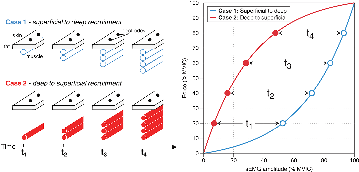
$p_i$ is the recorded motor-unit action-potential waveform, $a_i$ depends on electrode geometry and tissue conduction, $t_{ik}$ is spike timing, and $\eta(t)$ is noise. Recruitment changes $N$. Rate coding changes $K_i$ and $t_{ik}$. Electrode placement, subcutaneous tissue, fiber conduction velocity, cancellation, and movement change the recorded waveform.
The figure below shows two recruitment geometries that produce different relationships between force and surface EMG. This is why larger EMG amplitude cannot be translated directly into more recruited motor units.
Needle or fine-wire EMG samples individual units from a small tissue region. High-density surface EMG records many spatial channels, and decomposition algorithms estimate the discharge times of individual units. The next figure shows the progression from bipolar electrodes to a 64-channel grid and decomposed motor-unit spike trains.

Dynamic high-force lifting is harder to decompose than controlled isometrics because muscle fibers move relative to the electrodes and the signal changes w/ joint angle, velocity, and sweat on the measurement probes. Wearables that report an exact “activation percentage” from one surface sensor shouldn’t be treated as motor-unit counters, because it’s impossible to do accurately
10. voluntary recruitment and electrical stimulation aren’t identical
During voluntary contraction, synaptic input reaches motor-neuron cell bodies and generally follows the size principle. During peripheral electrical stimulation, the electric field depolarizes axons near the electrode. Recruitment depends on axon diameter, depth, orientation, electrode geometry, pulse width, and current.
Larger axons can have lower electrical thresholds, and surface stimulation can recruit units in a less orderly and more spatially fixed pattern than voluntary exercise. The same electrically recruited fibers may fire synchronously on every pulse, which produces force but can fatigue them quickly.
This is relevant to NMES after surgery, stroke, spinal-cord injury, or prolonged immobilization. Electrical stimulation can provide muscle loading when voluntary drive is limited, but its current, pulse width, electrode placement, and duty cycle determine which axons are reached and how fast fatigue develops.
11. what I would measure during lifting for the purpose of a scientific study
A useful set log can record:
exercise and ROM standard
load and repetitions
RIR
fastest and last-repetition velocity
rest time
pain location and intensity >> if abnormal
reason the set stopped
The basic calculations are:
$$ \text{volume load}=Wn $$
$$ \text{relative load} = \frac{W}{\widehat{1RM}} $$
$$ \text{velocity loss} = 1- \frac{v_{\mathrm{last}}} {v_{\mathrm{fastest}}} $$
These variables describe external work, relative intensity, fatigue, and remaining capacity more clearly than RPE alone. However a direct motor-unit research study requires IM EMG or validated high-density EMG decomposition under a controlled task.
TLDR. My personal learning: a motor unit is one alpha motor neuron and every muscle fiber supplied by its axon. The CNS increases force by recruiting more units and increasing the firing rates of active units. Heavy loads require high force immediately, while light loads can require high neural drive later as fatigue lowers available force. Plate weight reaches the motor-unit pool through acceleration, joint moment arms, muscle length, contraction velocity, and technique. Compound lifts can produce high whole-body RPE through bracing, breathing, grip, and coordination without proving maximal recruitment in one muscle. Caffeine can increase output without reliably lowering RPE, while creatine supports rapid ATP resynthesis rather than selecting motor units. In medicine, reduced recruitment can indicate motor-neuron or axonal loss, early recruitment can occur in myopathy, transmission can fail at the neuromuscular junction, and pain or joint effusion can inhibit voluntary drive. Surface EMG combines recruitment, firing rate, tissue conduction, electrode geometry, and noise, so it is not a direct motor-unit count.
Comments are closed for this post.